
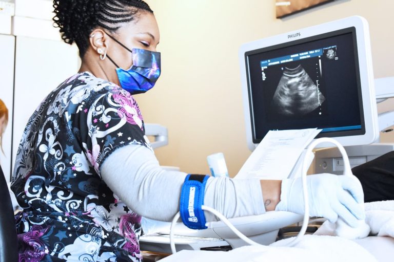
What Does a Diagnostic Medical Sonographer Do?
Sonography is the practice of using ultrasound imaging to non-invasively “view” structures inside the body with carefully directed, high-frequency sound waves.
A diagnostic medical sonographer carries out ultrasound procedures on patients and interprets the results. They prepare the equipment, perform the tests, and provide feedback for physicians and other medical professionals. They play an important role in helping assess the cardiac and circulatory health of patients.
The Purpose of Cardiac Ultrasound
Cardiac ultrasound is a type of ultrasound that examines blood flow through the patient’s arteries. It can be used to:
- Detect the presence of plaque
- Assess whether a patient is at risk of a stroke
- Determine whether the arteries have narrowed due to atherosclerosis
- Assess the condition of stents or outcomes of other medical interventions
Blood vessels may become narrowed if a plaque builds up inside them. These plaques restrict the flow of blood and can increase a person’s risk of a stroke or a transient ischemic attack (TIA). Medical professionals often recommend a cardiac ultrasound as a diagnostic procedure for people who have high blood pressure, high cholesterol, diabetes, or a family history of stroke or heart disease.
Cardiac ultrasounds are also used to detect hematomas (collections of clotted blood) following injuries, even in people who aren’t normally thought to be at risk of strokes.
How Do You Become a Medical Sonographer?
If you’re interested in becoming a medical sonographer, you’ll need to complete some extensive training. At Caris College, our sonographer training course leads to an Associate of Applied Science degree, including all of the qualifications required to work as a medical sonographer.
This accelerated 24-month program includes a 1,320-hour externship, giving trainees the hands-on experience they need to succeed in the field once they graduate. The training takes place in a fully functioning clinical lab, with training from instructors who have extensive real-world experience.
As part of the program, students will learn the practical skills they need to perform an ultrasound on the heart and blood vessels. At the end of the course, there’s the option to sit for the board exams required to move into the workplace as a cardiac sonographer.
If you’re interested in starting a new career or moving from a different area of healthcare into cardiac sonography, our sonographer training offers an accelerated route into the field. Discover more about Caris College’s cardiac sonography course and request an information pack or enroll today to take the first step towards a fresh and interesting new career.
Key Takeaways
What is a cardiac ultrasound?
A cardiac ultrasound is a non-invasive imaging test that uses sound waves to evaluate blood flow in the cardiac arteries and identify issues such as plaque buildup or narrowed arteries.
Why is a cardiac ultrasound performed?
It helps assess stroke risk, detect atherosclerosis, evaluate blood vessel health, and monitor the effectiveness of treatments such as stents.
Who performs a cardiac ultrasound?
Cardiac ultrasound procedures are typically done by diagnostic medical sonographers who have gone through a specialized training program like the one offered by Caris College.